
Diagnostics · in-house
Diagnostic centre
A full-fledged diagnostic centre inside the hospital: sonography with colour Doppler, obstetric and other ultrasound, ECG and NABL-accredited pathology, so reports come back quickly and treatment is never delayed.
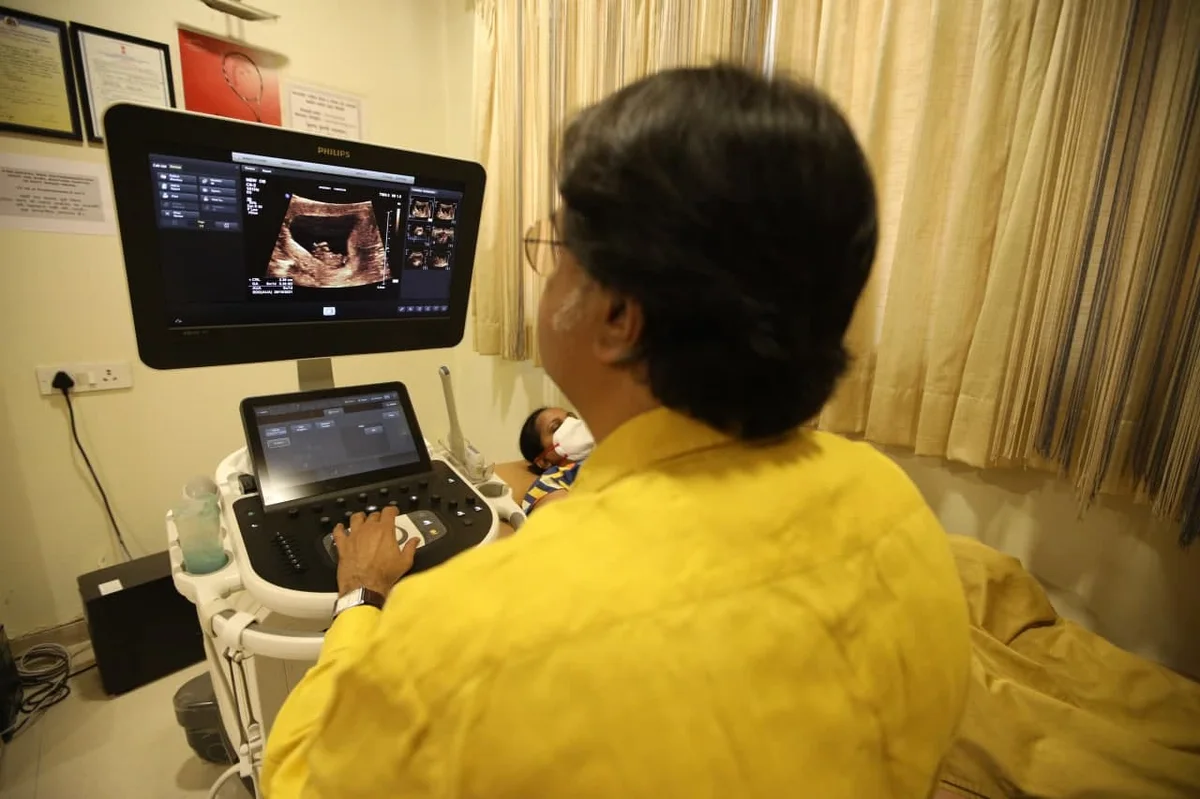
- SonographyPhilips HD 11 with 3D
- PathologyNABL-accredited lab
- Led byDr. Samir Vyas, radiologist
- SinceIn-house since 1985
Five services.
-
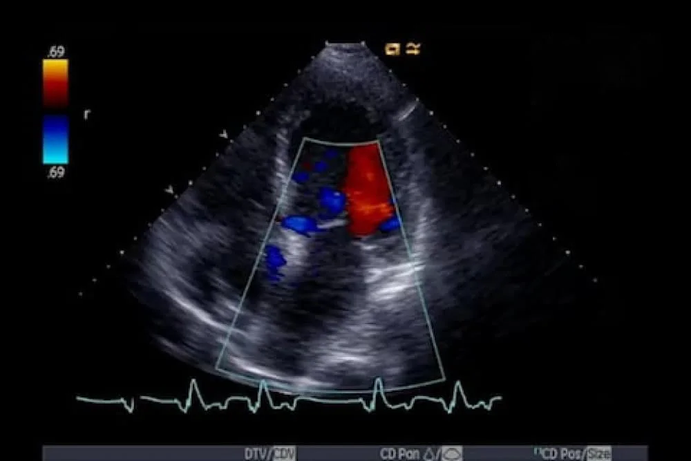
Colour Doppler studies
A colour Doppler ultrasound is a non-invasive procedure that visualises blood flow through blood vessels using high-frequency sound waves. A regular ultrasound uses sound waves to create images, but it cannot show blood flow. The check helps the doctor verify if you have a condition that is blocking your blood flow, as well as in diagnosing heart diseases. It is used in pregnancy to study blood circulation in the baby, uterus and placenta. Using it in high-risk pregnancies, where there is concern about the baby's condition, shows benefits.
-
Obstetric ultrasound
Obstetric ultrasound is a form of painless and safe ultrasound that does imaging of the foetus inside the uterus. It is also called ultrasound scanning or sonography. It uses a small probe called a transducer and gel placed directly on the skin. High-frequency sound waves travel from the probe through the gel into the body; the probe collects the sounds that bounce back, and a computer uses those sound waves to create an image. It provides reliable information during the pregnancy, such as determining the location of the pregnancy, the number of embryos present, and assisting in the identification of foetal anomalies.
-
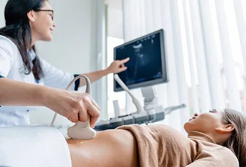
Other ultrasound
An ultrasound scan is a procedure that uses high-frequency sound waves to create images of organs and other internal structures. Ultrasound is used for screening of the abdominal and pelvic cavity, which involves the liver, gallbladder, pancreas, kidneys and bladder, in addition to obstetrics.
-
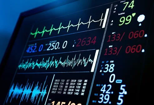
Electrocardiogram (ECG)
An electrocardiogram (ECG) is one of the simplest and quickest tests for recording the electrical activity of the heart. An electrical signal travels from the top to the bottom of your heart, triggering each heartbeat. This tracing is made up of representations of multiple waves that recur 60 to 100 times per minute with each heartbeat. The shape of the wave pattern should be consistent; if the waves are not consistent, this is indicative of a heart problem.
-
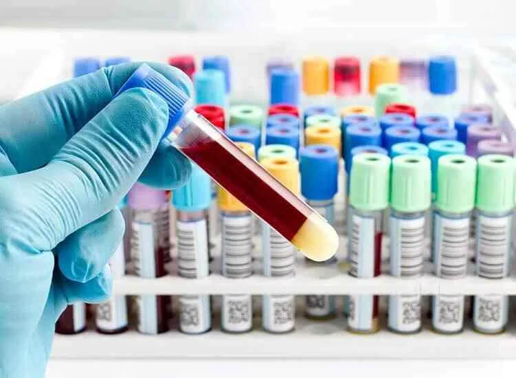
Pathology: full blood work
Pathology is a critical service in clinical diagnostics, and we have tied up with a NABL-accredited pathology lab to provide patients with effective care through error-free diagnosis. The lab is fully automated and fitted with cutting-edge technology. The lab is supervised by a highly trained pathologist, and the turnaround period for reports is short, which helps to reduce treatment delays.
Related care
-
Obstetrics & maternity
Antenatal care, normal, painless and C-section deliveries, high-risk pregnancy and antenatal exercises.
Learn more -

Gynaecology
Fibroids, PCOS, endometriosis, heavy bleeding, pelvic pain and laparoscopic surgery.
Learn more -
Well women clinic
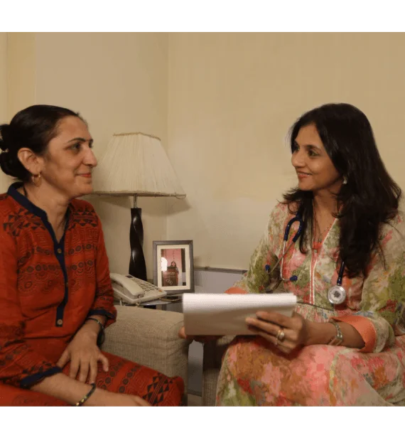
Health checks, breast and menopause care, sexual wellness advice and cancer screening.
Learn more
Book
Schedule a consultation call with us today.
Motherhood needs preparation. We can help.